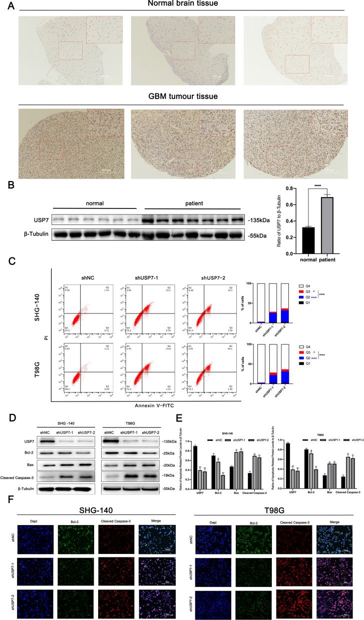
Fig. 1.
USP7 is highly expressed in GBM cells and its inhibition induces apoptosis. A Immunohistochemical staining of USP7 in human glioblastoma and normal brain tissue samples. Scale bar, 300 μm. B Western blotting analysis of USP7 protein levels in primary glioblastoma tissue samples and normal brain tissue samples, n > 6. C Apoptosis of SHG-140 and T98G cells treated by shUSP7s was measured by flow cytometry, n = 3. Q3 (Annexin V-FITC + PI-) subpopulation was considered early-stage apoptosis and Q2 (Annexin V-FITC + PI+) was late-stage apoptosis or necrosis. Cell proportion comparisons between NC and shUSP7 groups were done in both early- and late-stage subpopulation separately and in total. D, E. SHG-140 and T98G cells were treated with two different shUSP7s for 24 h. The changes in apoptotic proteins were observed by western blotting analysis, n = 3. F Immunofluorescence analysis of SHG-140 and T98G, cells were stained with DAPI and antibodies against BCL-2 or CLEAVED-CASPASE-3. Scale bar, 100 μm. Statistics are expressed as mean ± S.E.M., #P = NS, **P < 0.01 or ***P < 0.001

