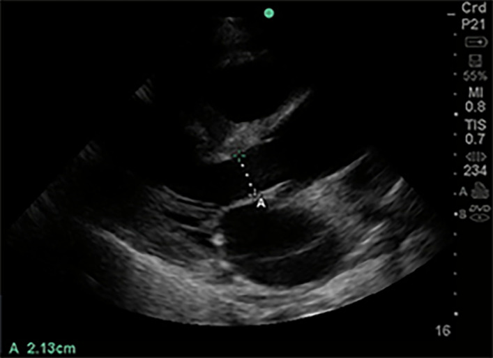
Figure 6.
Left ventricular outflow tract diameter parasternal long axis view. Of 1-5 MHz phased array probe with probe marker facing patient’s right shoulder, parasternal long axis view. Left ventricular outflow tract diameter measured during mid-systole, inner edge to inner edge, from septal endocardium to anterior mitral leaflet, in order to calculate cross-sectional area (πr2).

