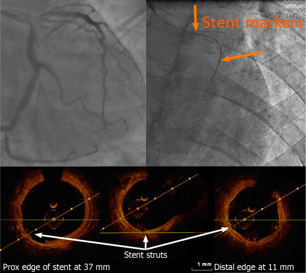
Figure 4.
Left anterior descending artery optical coherence tomography pull-back post bioresorbable stent implantation carried out using a 2.7 French imaging catheter with a dedicated workstation (C7 Dragonfly and C7-XR, Lightlab Imaging [Westford, Massachusetts, United States)]. Of note, only the proximal and distal markers are visible. On optical coherence tomography (OCT), the thickness of the stent struts and its good apposition can be assessed without artefact as seen with conventional metallic struts. Stent length could be measured on the OCT pullback with a good agreement with the known stent length.

