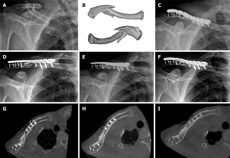
Figure 1.
Imaging examinations. A: Anteroposterior X-ray image of the initial injury; B: Three-dimensional computed tomography (CT) reconstruction images of the initial injury; C: Anteroposterior X-ray image after the internal fixation surgery; D: Anteroposterior X-ray image 3 mo postoperatively; E: Anteroposterior X-ray image 4 mo after shockwave therapy; F: Anteroposterior X-ray image 7 mo after shockwave therapy; G: CT scan image 3 mo postoperatively; H: CT scan image 4 mo after shockwave therapy; I: CT scan image 7 mo after shockwave therapy.

