Figure 4.

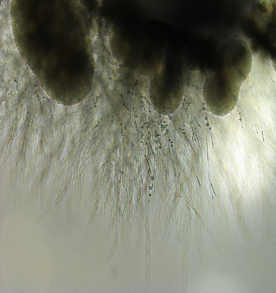
Appearance of Exophiala dermatitidis under stereoscopic microscope. Sputum sample of pneumonia patient from our institution. Melanized, dimorphic, dematiaceous, and hyphal-growth-state fungus, with multiple conidial forms, was confirmed.

Appearance of Exophiala dermatitidis under stereoscopic microscope. Sputum sample of pneumonia patient from our institution. Melanized, dimorphic, dematiaceous, and hyphal-growth-state fungus, with multiple conidial forms, was confirmed.