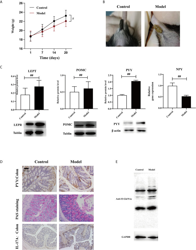
Figure 2.
Effects of HTHH conditions on appetite in mice exerted via the gut-brain axis (A) Body weight in the experimental group was significantly lower than that in the control group. (B) The appearance and morphology of feces differed in the control group and the experimental group. (C) Western blotting results of LEPT and POMC expression in the hypothalamus, and PYY expression in the colon. NPY expression in the hypothalamus was detected via qPCR (n = 3, two-tailed Student’s t-test. #p < 0.05, ##p < 0.01. (D) PYY and IL-17A expression detected via immunohistochemistry. Periodic acid–Schiff staining was used to detect glycogen and mucin in colon tissue. (E) GlcNAcylation in the colon was detected using a pan anti-O-GlcNAc monoclonal antibody (n = 3).

