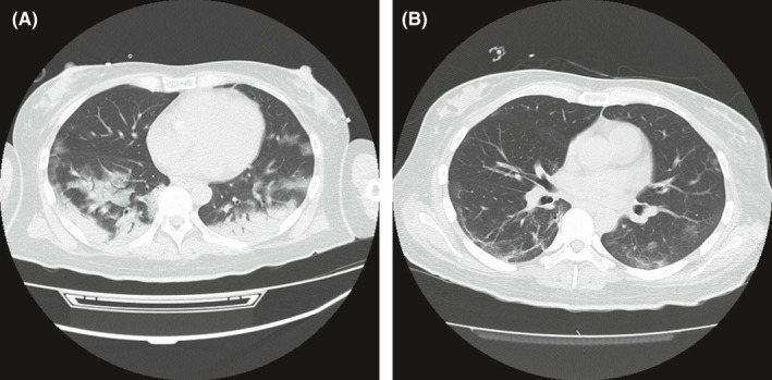
FIGURE 1.

(A) Chest computed tomography on Day 1 of hospitalization showed bilateral distribution of GGO with consolidation in the posterior lobe and peripherally including crazy paving, air bronchogram, and a reticular pattern. (B) Improving bilateral GGO and fibrous stripes were observed in the lower lobes on Day 15. GGO, ground‐glass opacities
