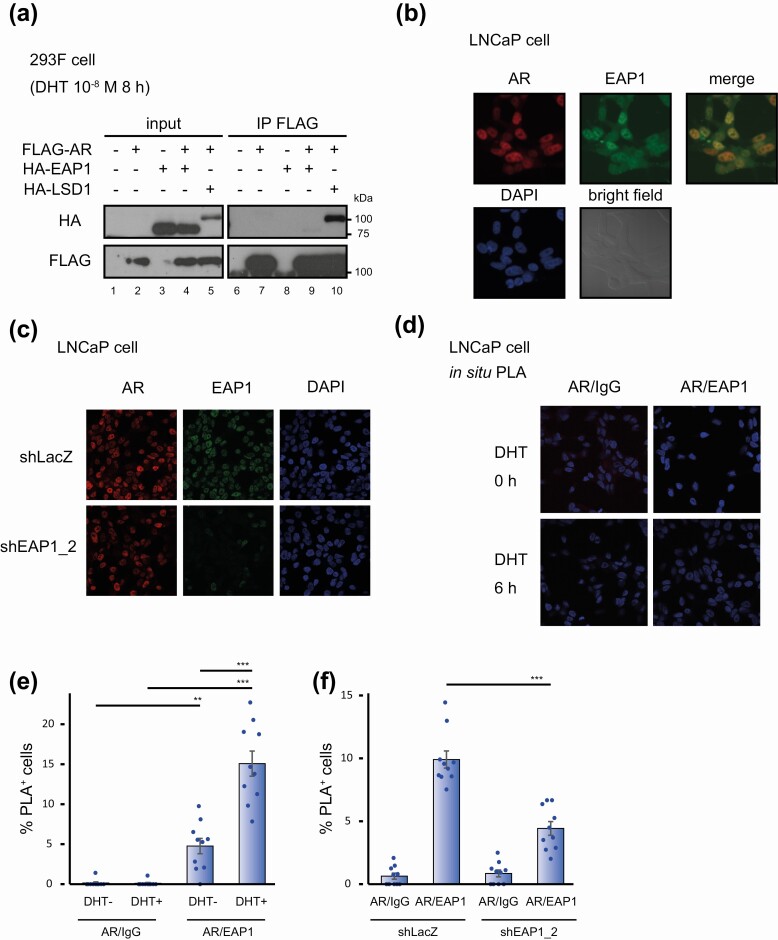
Figure 2.
Androgen receptor (AR) interacts with enhanced at puberty 1 (EAP1) in the cells to a degree that it could not be detected under the standard biochemical condition. A, 293F cell lysates exogenously expressing FLAG-tagged AR and/or hemagglutinin (HA)-tagged EAP1 and stimulated with 10-nM dihydrotestosterone (DHT) for 8 hours were subjected to immunoprecipitation followed by immunoblot analysis using anti-FLAG and anti-HA antibodies. HA-tagged lysine-specific demethylase 1 (LSD1) was used as a positive control for coimmunoprecipitation. The molecular weights of the marker proteins are indicated on the right. B, LNCaP cells treated with 10-nM DHT for 6 hours were stained with anti-AR and anti-EAP1 antibodies and 4′,6-diamidino-2-phenylindole (DAPI). Both antigens were colocalized in the nucleus of the cells. Bright field images are also shown. C, LNCaP cells that expressed short hairpin RNA (shRNA) for EAP1 were treated with 10-nM DHT for 6 hours and stained with anti-AR and anti-EAP1. LNCaP cells expressing shRNA for LacZ were used as a control. As for EAP1 expression levels in these cells, see Fig. 3C and 3D. D, LNCaP cells were treated for 6 hours with vehicle (ethanol) or with 10-nM DHT and were analyzed by proximity ligation assay (PLA) to validate protein interactions between AR and EAP1. Images were acquired at 40× magnification. E, Quantification of the number of PLA dot–positive cells in Fig. 1D. Data were analyzed by one-way analysis of variance (ANOVA) with a post hoc Tukey-Kramer test (n = 10) Error bars indicate SDs. *P less than .05; **P less than .01; ***P less than .001. F, PLA was performed using 10-nM DHT–treated LNCaP cells (6 hours) that expressed either shLacZ or shEAP1. Quantification of the number of PLA dot–positive cells are shown. Data were analyzed by one-way ANOVA, with a post hoc Tukey-Kramer test (n = 10) Error bars indicate SDs. ***P less than .001.

