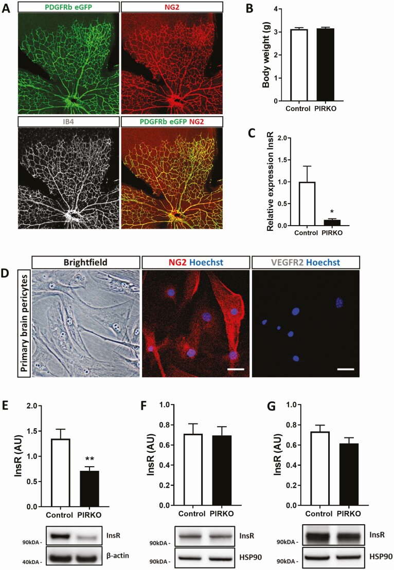
Figure 2.
Confirmation of appropriate pericyte insulin receptor knockout. (A) Cre-recombination was assessed in PDGFRβ-CremTmG+/– mice. Endogenous eGFP expression colocalizes with pericyte NG2 expression in P5 retinas. The vasculature was labeled with isolectin B4 (gray). (B) There is no difference in body weight at P5 between PIRKO and control. (C) Brain pericytes were isolated from adult control and PIRKO mice. Knockdown of the insulin receptor was confirmed on RNA level (relative to β-actin). (D) Isolated brain pericytes at passage 5 express the pericyte marker NG2 (central panel), but not the endothelial marker VEGFR2 (right panel), scale bar 50 µm. (E) Insulin receptor protein level in reduced in isolated brain pericytes from PIRKO. (F) PDGFRβ-targeted insulin receptor knockdown does not affect whole tissue insulin receptor expression in P5 retinas and (G) P5 lungs. Data presented as mean ± SEM, unpaired t-test, *P < .05, **P < .01, n = 6, 6 (C), n = 9, 9 (E), n = 7, 7 (F,G).

