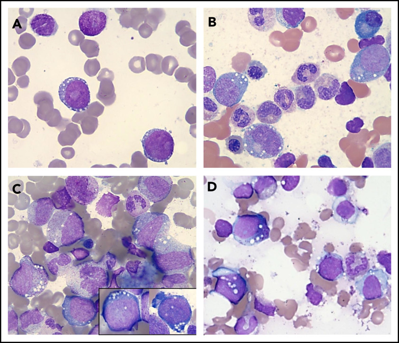
Figure 1.
Vacuolization of HPs in various conditions. Wright-Giemsa stains (magnification ×1000) showing a representative example of a "blast-only” pattern of vacuolization in a case with high-risk MDS (vacuoles in 2 blasts) (A) and vacuolization of immature erythroid and myeloid cells in 2 patients with a UBA1 mutation and VEXAS syndrome (B-C) and in a patient with copper deficiency (D).

