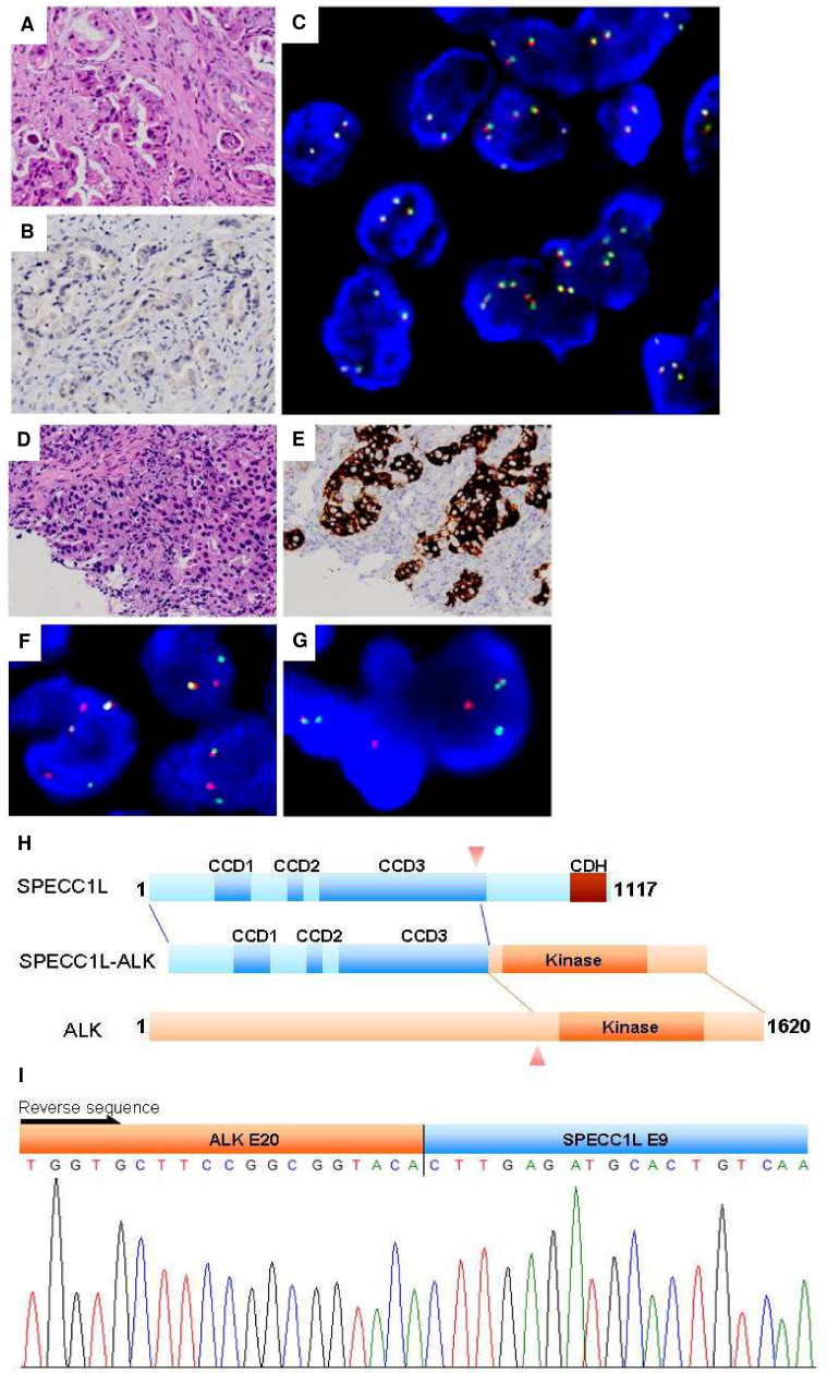
Fig 3. Analysis of ALK status by ALK KD screening, IHC, and FISH.
FFPE discrepancy cases. Case E1154 brain metastasis: (A) H&E stain, (B) ALK IHC, (C) ALK break-apart probes. No break-apart event was identified. Many cells showed more than 2 fused signals. Case E1757 SPECC1L-ALK: (D) H&E stain, (E) ALK IHC, (F) ALK break-apart probes, (G) SPECC1L-ALK probe in tumor cell containing the SPECC1L-ALK fusion (one orange/green (yellow) fusion signal was observed), (H) schematic representation of the SPECC1L-ALK protein, (I) 5’-RACE and sequencing confirmed.

