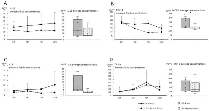
Fig 3. Cytokines/chemokine concentrations in amniotic fluid after the administration of either saline or rytvela).
Cytokines/chemokine concentration in AF at each time point plotted as mean ± SD. Each group’s average concentration is shown in bar charts. A, IL-1β; B, MCP-1; C, IL-8; D, TNF-α. The average concentration was compared between the LPS Group and the LPS + rytvela Group. *p < .05 between the LPS Group and the LPS + rytvela Group. n = 3–7 animals/group. AF, amniotic fluid; SD, standard deviation; IL, Interleukin; MCP, monocyte chemoattractant protein; TNF, tumor necrosis factor.

