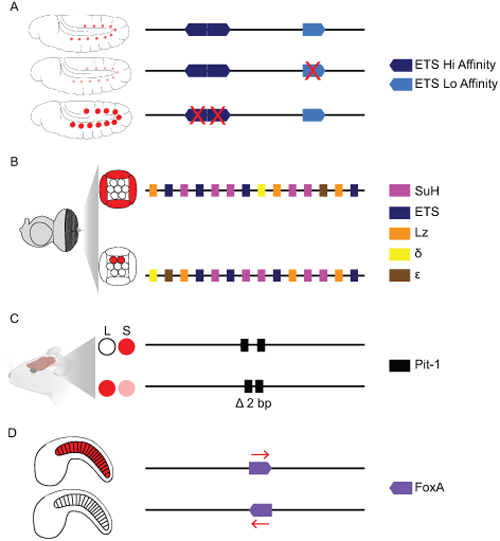
Figure 3: Affinity, Order, Spacing, and Orientation of sites within enhancers are essential for encoding tissue-specific gene expression during development.

(A) A combination of low-affinity and high-affinity ETS sites within the MHE enhancer restrict expression to the muscle and heart cells of the developing fly. Low-affinity sites bind an activator form of ETS called Pointed, removing low-affinity sites reduces expression. High-affinity sites bind a repressive form of ETS called Yan, loss of high-affinity sites leads to ectopic expression (Boisclair Lachance et al., 2018). (B) In the developing fly eye, specific orders of SuH, ETS, LZ, δ and ε TFBSs activate expression in the cone cells and repress expression in the photoreceptor cells. Changing the order of sites can lead to loss of expression in the cone cells and ectopic expression in the photoreceptor cells (Swanson et al., 2010). (C) In the developing mouse pituitary, a 2-bp spacing within a Pit-1 site allows for expression in the somatotrope (S) cells and repression in lactotrope (L) cells, removing this spacing leads to ectopic expression of GH in lactotrope cells (Scully et al., 2000). (D) In Ciona, the orientation of a FoxA site within the Tune enhancer ensures notochord expression (Passamaneck et al., 2009). Not all sites within each enhancer are shown; for simplicity, we focus on the sites that when manipulated impact gene expression. Expression patterns are shown in red.
