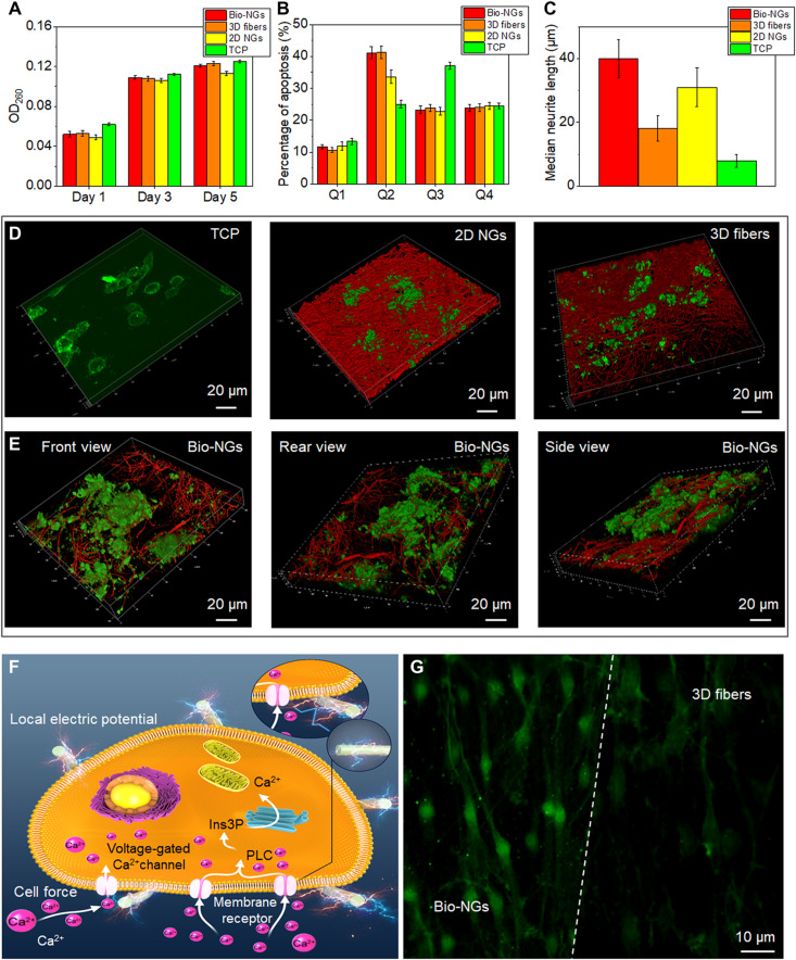
Fig. 4. The growth and development of RGC5 neurons in bio-NGs.
(A) Proliferation of RGC5 neurons by the DNA assay on days 1, 3, and 5. (B) Apoptosis of RGC5 neurons after 5 days of culture in bio-NGs. (C) Neurite outgrowth of RGC5 neurons by the median neurite length after 5 days of culture in bio-NGs. (D) 3D confocal scanning of RGC5 neurons cultured on TCP, 2D NGs, and 3D fibers. (E) 3D confocal scanning of RGC5 neurons cultured in bio-NGs from different perspectives. (F) Inherent cell force of living cells grown in bio-NGs. This would induce a local electrical field proportional to the strain level that could eventually alter the membrane potential and/or the configuration of membrane receptors and results in the opening of the Ca2+ channels. Ins3P, inositol trisphosphate. PLC, phospholipase C. (G) The fluorescence images of the cells preincubated with Fluo-4 AM (membrane-permeable and Ca2+-dependent dye) on the fibers in bio-NGs and 3D fibers. Green, Ca2+. All error bars indicate ±SD.

