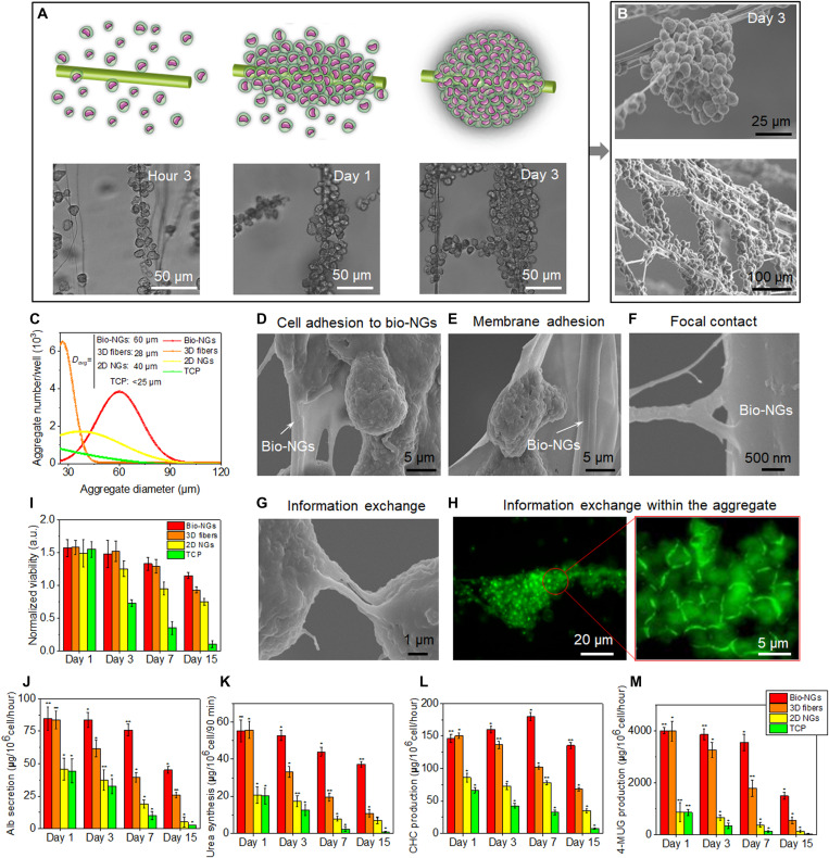
Fig. 5. The motility and function maintenance of primary hepatocytes in bio-NGs.
(A) Schematic diagram and light microscope images of hepatocyte movement to form cell clusters. (B) SEM images of hepatocyte aggregates. (C) Hepatocyte aggregate size and number on day 3. (D to F) Cell morphology and NG-cell interaction including cell membrane adhesion and focal contact, assessed by SEM after 15 days in culture, showed that hepatocytes were adhered to fibers in bio-NGs. (G) SEM image showing the information exchange between cell aggregates. (H) Laser scanning confocal microscopy images of epithelial cadherin (green) detection showing the cellular information exchange within cell aggregates. (I) MTT assay showing no signs of toxicity for cells cultured in bio-NGs. Hepatic function assessment: (J) Albumin (Alb) secretion and (K) urea synthesis at different culture time points. The metabolic functions of hepatocytes: (L) 3-cyano-7-hydroxycoumarin (CHC) and (M) 4-methylumbelliferyl glucuronide (4-MUG) production at different culture time points. All error bars indicate ±SD. n = 3. *P < 0.05 and **P < 0.01.

