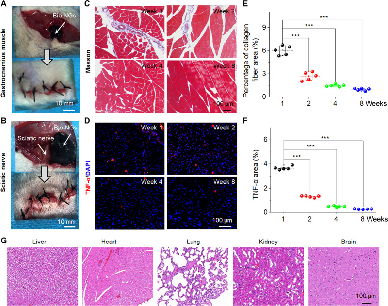
Fig. 7. In vivo stability and biocompatibility of bio-NGs.
Surgical image showing the implantation of the bio-NGs into the (A) gastrocnemius muscle and (B) sciatic nerve areas of a mouse. (C) Masson trichrome staining of gastrocnemius muscles at the implanted area. (D) TNF-α immunofluorescent staining of sciatic nerve at the implanted area. (E) Average percentage of collagen fibers in the muscle tissue measured from Masson staining. (F) Relative TNF-α expression level measured from TNF-α immunofluorescent staining. (G) H&E staining of vital organs (liver, heart, lung, kidney, and brain) at week 8 after implantation in the sciatic nerve area. Data are expressed as mean values ± SD. n = 5. ***P < 0.001. Photo credit: Tong Li, Nanjing University of Science and Technology.

