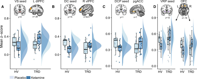
Fig. 1. Group differences in the effects of ketamine on functional connectivity across four striatal seeds.
Ketamine differentially altered functional connectivity between the groups, as reflected in VS-left dlPFC (a), DC-right vlPFC (b), DCP-pgACC (c), and VRP-left/right OFC (d) coupling. This was identified using group-by-treatment F–tests at a family-wise error (FWE) cluster-corrected threshold level of p < 0.05. Boxplots with individual data points and distributions [75] show that functional connectivity was increased in individuals with treatment-resistant depresssion (TRD) but reduced in healthy volunteers (HVs) post-ketamine relative to placebo (a–d). Resting-state functional magnetic resonance imagining scans (rsfMRI) were acquired 2 days after each infusion. VS ventral striatum; DC dorsal caudate; DCP dorsal caudal putamen; VRP ventral rostral putamen; dlPFC dorsolateral prefrontal cortex; vlPFC ventrolateral prefrontal cortex; pgACC perigenual anterior cingulate cortex; OFC orbitofrontal cortex; L left; R right; FWE family–wise error.

