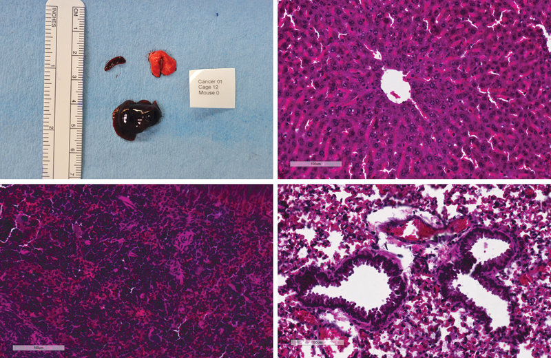
Figure 6:
Metastasis screening. Macroscopic aspect of liver, lung and spleen specimens collected from one animal during necropsy. No macroscopic metastasis was identified (a). Histological screening for metastasis was performed on liver (b); spleen (c); and lung (d). No microscopic metastasis was identified in HE slides. Ruler on macroscopic picture correspond to centimeters. Scale bars on HE microscopic pictures correspond to 100um.

