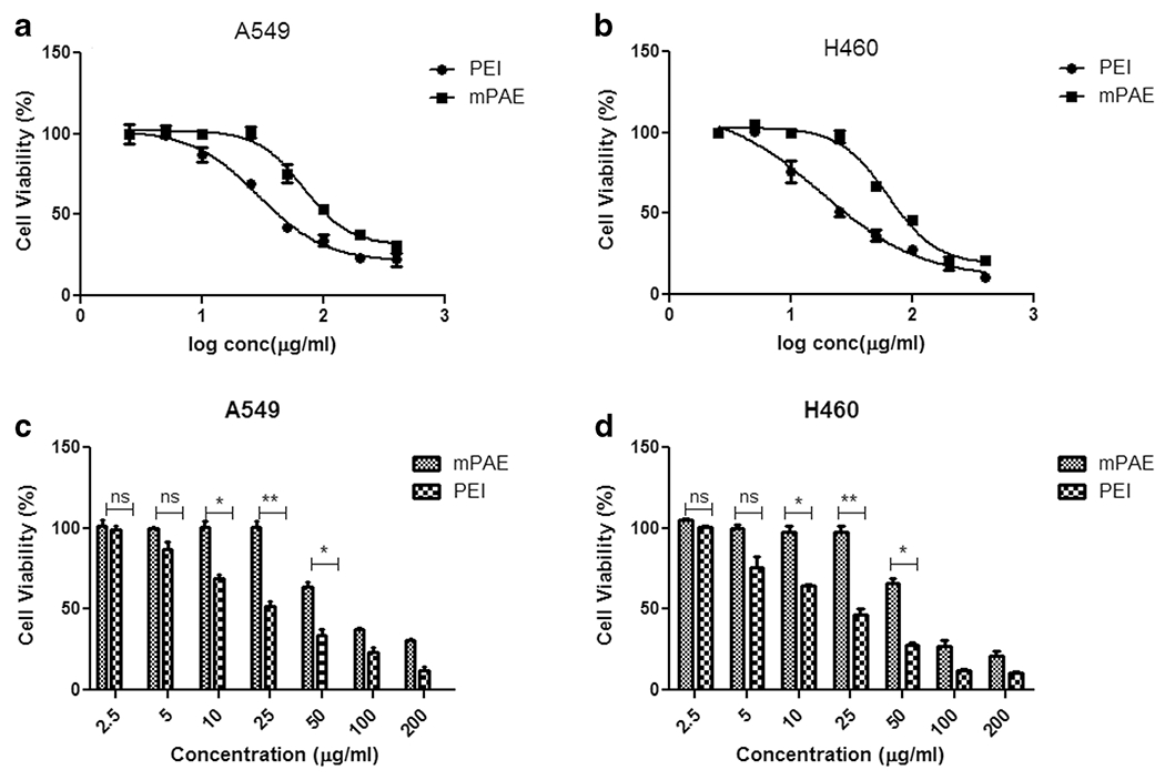
Fig. 3. Cytotoxicity analysis of mPAE and PEI polymers.

(a) Cell viability (%) of “mPAE and PEI” polymers at various log concentrations in (μg/ml) A549 cell line (b) Cell viability (%) of “mPAE and PEI” polymers at various log concentrations (μ/ml) in H460 cell line (c) Cell viability (%) after treatment of mPAE and PEI at concentrations (2.5, 5, 10, 25, 50, 100 and 200 μg/ml) in A549 cell line. and (d) Cell viability (%) after treatment of mPAE and PEI at concentrations (2.5, 5, 10, 25, 50, 100 and 200 μg/ml) in H460 cell lines. The cytotoxicity was determined using SRB assay after 48 h polymer exposure (n = 3, error bars represent standard deviation).
