. 2021 Sep 24;54:e0469-2021. doi: 10.1590/0037-8682-0469-2021

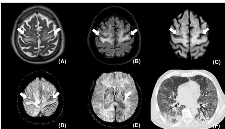
FIGURE 1: Axial T2 (A), FLAIR (B), and diffusion (C) weighted images demonstrating symmetric diffuse hyperintensity (arrows) and mild restricted diffusion (arrows) involving the subcortical white matter in the precentral gyrus and the posterior portions of the superior and middle frontal gyrus. Axial susceptibility-weighted images, (D) and (E), depict punctate microhemorrhagic foci on the cortical surface of the precentral gyrus, middle frontal gyrus, and superior frontal gyrus (arrows in D) and in the posterior limb of the internal capsule (arrow in E). Chest CT, pulmonary window, axial: diffuse areas of ground-glass opacities and consolidations (F).

FIGURE 1: