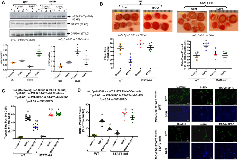
Figure 2.
Role of STAT3 in hearts of diabetic mice following rapamycin treatment. (A) Representative immunoblots for p-STAT3, total STAT3, and GAPDH expression in whole heart of C57 and db/db mice following 28 days of RAPA treatment. Densitometry analysis of immunoblots for the ratio of p-STAT3/STAT3 (*P < 0.05 vs. others) and the ratio of STAT3/GAPDH (*P < 0.05 vs. C57-Control; n = 3). (B) Myocardial infarct size following ischaemia/reperfusion in STAT3-deficient diabetic mice. High-fat diet (HFD)-fed WT and STAT3-deficient mice were treated with RAPA for 28 days prior to evaluation of I/R injury. Upper panel: representative images of heart sections following TTC staining (Scale indicates 5 mm). Lower panels: quantitative data of infarct size following I/R injury (*P < 0.001 vs. other) and rate-force product (*P < 0.01 vs. other; n = 5). (C) Isolated cardiomyocyte necrosis was determined by trypan blue staining following 40 min SI and 1 h RO (n = 4–8). (D) Representative pictures of TUNEL staining (scale indicates 100 µm) and quantitative data of cardiomyocyte apoptosis following 40 min SI and 18 h RO (n = 4; *P < 0.001 vs. controls, αP < 0.001 vs. both SI/RO and βP < 0.05 vs. WT-SI/RO). Statistics: one-way ANOVA.

