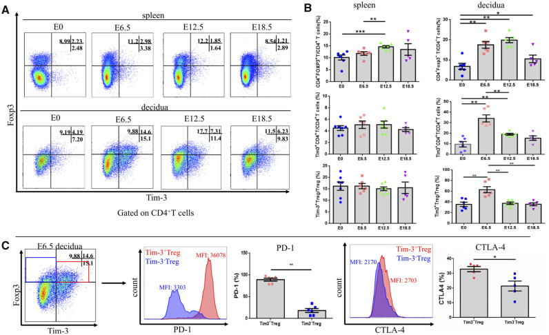
Figure 1.
Tim3+ Treg cells accumulated in the decidua of mice during early pregnancy. Tim-3 expression on CD4+ T cells and Treg cells in spleen and decidua at time points throughout pregnancy was determined by flow cytometry (n ≥ 6 mice/group). (A) Representative Tim-3 expression on spleen and decidual Treg cells gated on CD4+ T cells. (B) Statistical analysis of Treg cell, Tim-3-expressing CD4+ T cell and Tim-3-expressing Treg cell proportions in the spleen and decidua throughout pregnancy. (C) The frequency and mean fluorescence intensity (MFI) of PD-1 and CTLA-4 expression were assessed and compared between Tim-3+ Treg cells and Tim-3– Treg cells from the decidua on E 6.5. Data are represented as means ± SEM. *P < 0.05, **P < 0.01, ***P < 0.001. E, embryonic day; detection of vaginal plug = E 0.5.

