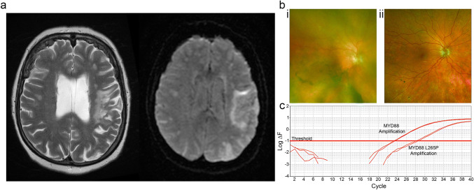
Fig. 3.
Diagnosis of primary CNS lymphoma with vitreous biopsy. a Patient presented with right-sided weakness with stuttering progression over several months. Left panel, T2-weighted MRI, increased signal in the territory of the left middle cerebral artery. Right panel, Diffusion-weighted imaging, increased signal suggesting restricted diffusion in left-hemisphere white matter. b Photographs of right fundus for patient in a: (i) before vitreous biopsy, retina obscured by cloudy vitreous; (ii) post-vitreous biopsy, retina visible, no sub-retinal deposits identified. c Detection of MYD88 L265P mutation by allele-specific PCR. The test compares the patient’s DNA with a positive control that has MYD88 L265P at 0.625%. The test relies on a failure of extension of the primers when there is mismatch between the primer and the extracted DNA. Any difference between the patient’s sample and the positive control is amplified by repeated PCR cycles and is measured (ΔF). The presence of the MYD88 L265P mutation is reported when the difference crosses a threshold

