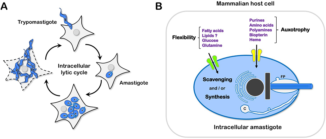
Figure 1.
(A) Trypanosoma cruzi infection cycle in mammalian host cells. Motile T. cruzi trypomastigotes actively invade nucleated mammalian cells to establish intracellular infection. Host cell entry triggers a developmental program [15], resulting in the formation of intracellular amastigotes that coincides with the localization of the parasite in the host cell cytoplasm. The timing of this process varies with parasite strain and host cell type. Replication competent amastigotes are typically formed by 16–20 hours post-infection (hpi) and begin to proliferate at ~24 hpi [27]. After several rounds of replication and cell division, intracellular amastigotes cease division and differentiate to trypomastigotes that eventually egress from the host cell where they can invade other cells. (B) Interaction of intracellular T. cruzi amastigotes with host nutrient sources. Generalized model depicting the transport of nutrients for which amastigotes have strict dependence on the host cell (auxotrophy) and those that can be acquired and synthesized by the parasite (flexibility). Transporters are drawn on the plasma membrane for simplicity but can be distributed within the flagellar pocket (FP) and cytostome/cytopharynx (C), and other endocytic organelles.

