Figure 4.

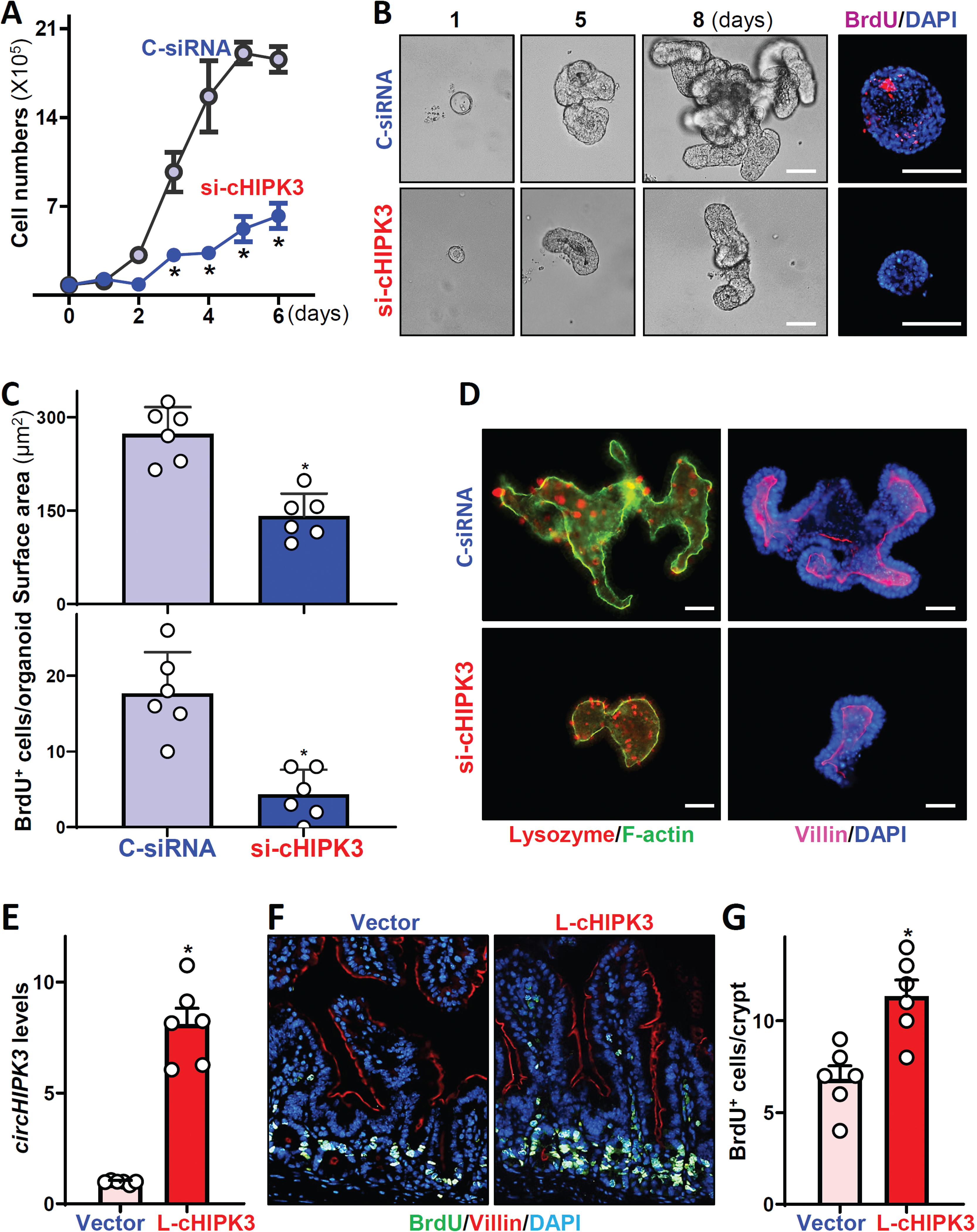
CircHIPK3 is essential for renewal of the intestinal epithelium. (A) Cell growth after circHIPK3 silencing in vitro. Values are means ± SEM (n = 3). * P < 0.05 compared with C-siRNA. (B) Growth of small intestinal organoids after circHIPK3 silencing ex vivo. Left: bright field microscopy analysis of growth of organoids on day 8; and right: confocal analysis of BrdU (red) and DAPI (blue) on day 3 after culture. Scale bars: 100 μm. (C) Quantification of surface area (top) and BrdU positive cells (bottom) of the organoids described in B (n = 6). (D) Immunostainings of lysozyme- (left) and villin-positive (right) cells in intestinal organoids on day 5 after transfection with si-cHIPK3 or C-siRNA. Red, lysozyme; purple, villin. Scale bars: 100 μm. (E) Levels of circHIPK3 in the small intestinal mucosa of mice on day 5 after intraperitoneal injection with a recombinant circHIPK3 lentiviral expression vector (L-cHIPK3) or empty control lentiviral vector (Vector). * P < 0.05 compared with control vector (n = 6). (F) Proliferating cells in small intestinal crypts as measured by BrdU labeling (1 h post-injection, green) in mice described in E. (G) Quantification of BrdU-positive cells in the mucosa described in F. * P < 0.05 compared with control vector (n = 6).
