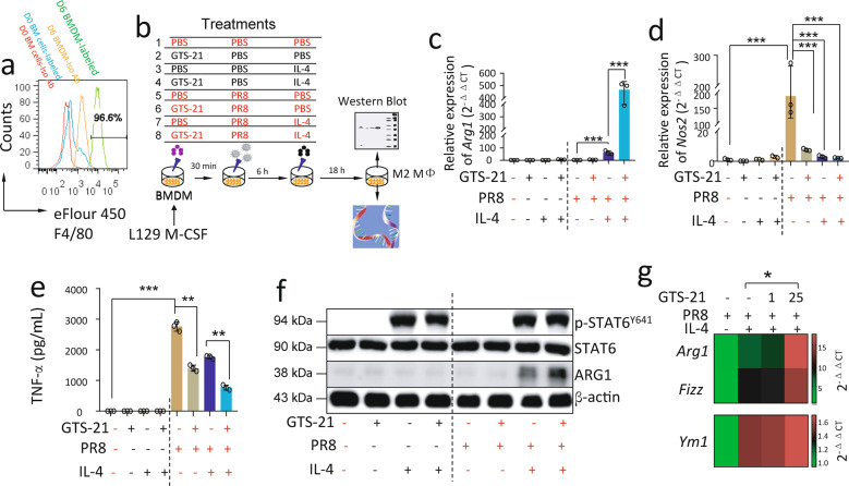
Fig. 4. Activation of α7nAChR enhances M2 MΦ activation in IL-4-treated PR8-infected BMDM.
a Histogram of F4/80 expression in BMDM. BMDM were collected after 6 days of culture and stained with fluorescent isotype or anti-F4/80 antibodies. The freshly isolated bone marrow cells were used as control. Experiments were repeated 2 times. b The experimental conditions for inducing M2 MΦ activation. Eight treatment strategies were selected, while the 8th strategy was as follows: BMDM (5 × 10⁵/well) received α7nAChR activation with GTS-21 (25 μM in PBS) for 30 min, IL-4 stimulation (10 ng/mL in PBS) for 6 h, and then PR8 (2 MOI in PBS) influenza challenge for 18 h. The other strategies were used as controls (the same concentration of cell, GTS-21, or IL-4 as the 8th was added as required). The cells were collected after 24 h treatment to extract RNA and protein for analysis. c–e Relative expression of Arg1, Nos2, and TNF-α expressions in-PR8-infected BMDM treated with C8 condition for 18 h. The cells were collected to extract RNA for measuring Arg1 and Nos2 expression. Supernatants of media were collected to measure TNF-α by ELISA (e). n = 3 in each group. **P < 0.01, ***P < 0.001 in the indicated groups, One-way ANOVA. Cells were collected to measure p-STAT6 and ARG1 in cell lysates by Western blot (f). g Relative expression of Arg1, Fizz, and Ym1 mRNAs in α7nAChR-activated IL-4-induced PR8-infected BMDM. n = 3 in each group, *P < 0.05, unpaired t test. GTS-21 concentration was used from 0, 1, and 25 μM. Using strategies listed in (b). Data in (c–g) were representatives of two independent experiments. “-” condition indicated PBS treatment.

