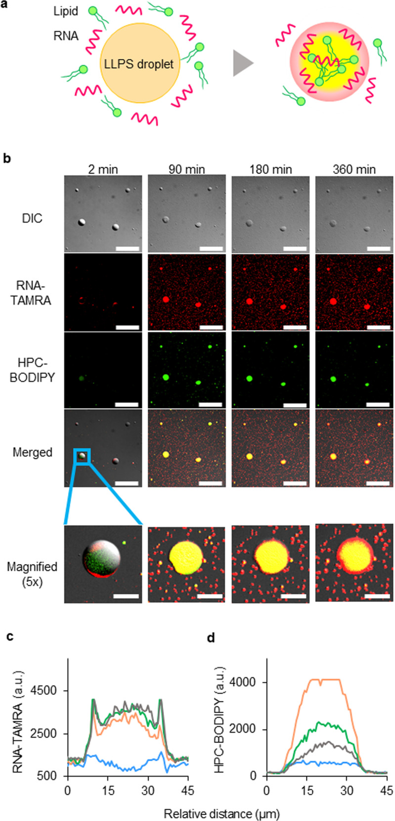
Fig. 6. Incorporation, concentration, and separation of biological molecules inside LLPS-formed droplets by internal structural heterogeneity. Nucleic acids and lipids that were concentrated in the LLPS-formed droplets exhibited higher-order functions of the droplets.

a Due to the spatial heterogeneity of a LLPS-formed droplet, the RNA and lipids were concentrated and spatially separated, and higher-order properties that had not been exhibited by the droplet itself were apparent. b Confocal laser scanning (CLS) fluorescence microscopy images of the LLPS-formed droplet dispersion at 2, 90, 180, and 360 min after addition of the TAMRA-RNA (10 μM) and BODIPY-HPC (10 μM) solutions; both the individual and merged signals are shown (DIC = differential interference contrast microscopy image). The scale bars of the images in the top four rows are 100 μm, whereas those in the bottom row (magnified) images represent 20 μm. The blue rectangle in the photo in the merged image is a magnification of the image below. The result was verified by five trials. c, d Temporal evolution of line profiles of fluorescence intensities along a horizontal line through the centre of the LLPS-formed droplet after addition of TAMRA-RNA and BODIPY-HPC: blue line, 2 min; yellow line, 90 min; green line, 180 min; black line, 360 min. c Line profiles of TAMRA-RNA fluorescence intensity of the LLPS-formed droplet shown in the bottom row of (b). d Line profiles of BODIPY-HPC fluorescence intensity of the LLPS-formed droplet shown in the bottom row of (b). RNase-free water was used as a substitute for deionised water. Source Data of Fig. 6c, d are provided in the Source Data File.
