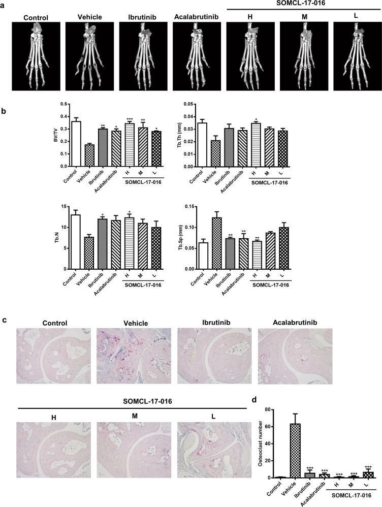
Fig. 3. SOMCL-17-016 hampered bone erosion and diminished excessive osteoclasts in CIA mice.
a Representative three-dimensional reconstructions of the right hind paws of normal control mice and CIA mice. b Histomorphometric analysis of the distal tibia for each treatment group and the bone volume/total volume (BV/TV), trabecular thickness (Tb. Th), trabecular number (Tb. N), and trabecular spacing (Tb. Sp) values of mice from each group. c TRAP staining (magnification of ×100) of representative inflamed joints in the hind paws of mice from the different groups. d Numbers of TRAP-positive osteoclast cells in a field of joint sections of mice from each group. The data are shown as the means ± SEMs (n = 3); *P < 0.05, **P < 0.01, and ***P < 0.001, significantly different from the vehicle-treated group.

