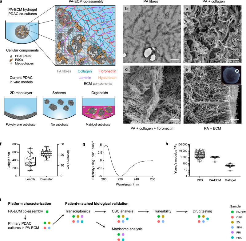
Fig. 1. Design and characterisation of PA-ECM co-assembling matrices.
a Schematic illustration of PA hydrogel co-assembly with ECM macromolecules compared to currently used substrates for the in vitro modelling of PDAC. b Transmission electron micrograph of PA fibres. Scale bar: 100 µm. c–e Scanning electron micrographs of PA hydrogels co-assembled with collagen (c), collagen and fibronectin (d) and all ECM components (e); insert indicates imaged area within a 2 µL hydrogel. Scale bar: 2 µm. Insert scale bar: 500 µm. f PA fibre size measured from TEM micrographs. Box plots indicate range, interquartile range and median. n = 20 individual fibres. g PA fibre circular dichroism spectrum in HEPES buffer. h Stiffness (Young’s modulus) of PDX tissue, as measured by atomic force microscopy (n = 71 measurements across a tissue section), compared to PA-ECM (n = 24 measurements of a 10 mg/mL gel) and Matrigel (n = 38 measurements of a 10 mg/mL gel), as measured by rheometry. Box plots indicate range, interquartile range and median. i Experimental outline for the validation of the platform as a model for pancreatic cancer.

