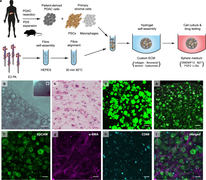
Fig. 2. PA-ECM cultures for the ex vivo modelling of pancreatic cancer.
a Schematic illustration of 3D cell culture in PA-ECM. b Brightfield micrograph of PDAC cells co-cultured with PSCs and macrophages in PA-ECM; insert indicates imaged area within a 5 µL hydrogel. Scale bar: 50 µm. Insert scale bar: 500 µm. c Haematoxylin (blue) and eosin (red) stain of PA-ECM hydrogel triple culture. Scale bar: 50 µm. d 3D projection of PDAC cells grown in PA-ECM for 14 days. Living cells were stained with calcein AM (green) and dead cells with ethidium homodimer (red). Scale bar: 100 µm. e 3D projection of PDAC cells co-cultured with PSCs and macrophages in PA-ECM hydrogels for 7 days and stained for EpCAM (green) and Ki-67 (white). Scale bar: 100 µm. f–i 3D projection of a PA-ECM hydrogel triple culture. PDAC cells were identified by EpCAM (green)(f), PSCs by α-SMA (magenta) (g) and macrophages by CD68 (cyan) (h) immunostaining. All cell types are shown on (i). Scale bars: 50 µm.

