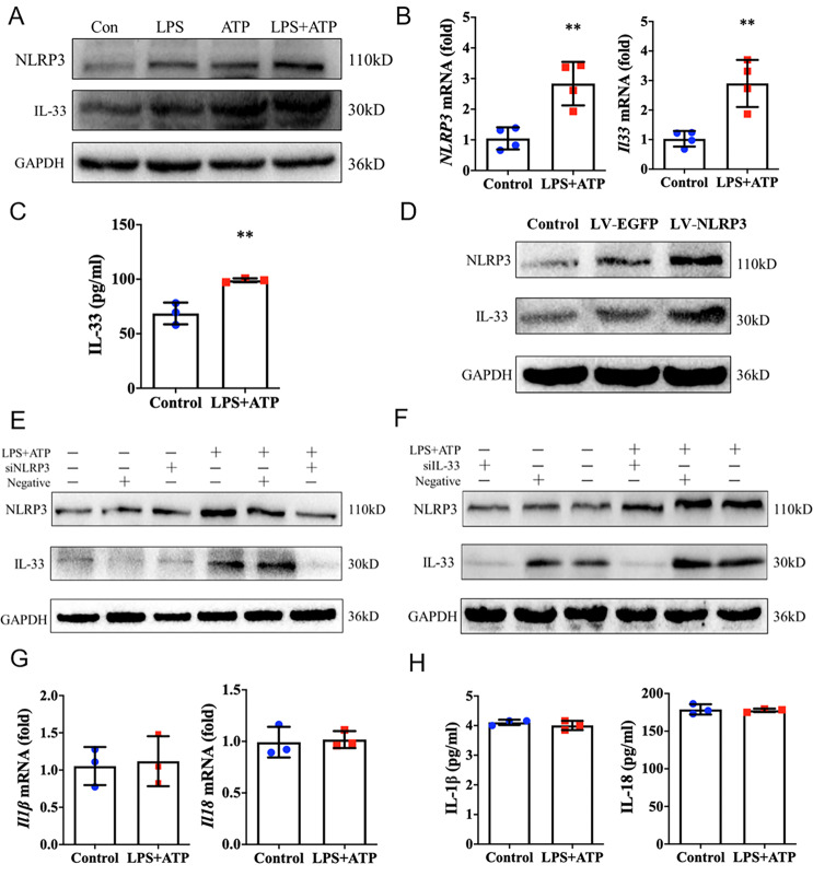
Fig. 2. NLRP3 regulates IL-33 expression and secretion in epithelial cells.
A Immunoblot analysis of NLRP3 and IL-33 protein expression in HaCaT cells in response to LPS (10 μg/mL, 24 h), ATP (5 mM, 1 h), or LPS (10 μg/mL, 24 h) and ATP (5 mM, 1 h) stimulation. B Relative expression of Nlrp3 and Il33 mRNA in HaCaT cells in response to LPS (10 μg/mL, 24 h) and ATP (5 mM, 1 h) stimulation by qRT-PCR analysis. C ELISA analysis of IL-33 secretion level from HaCaT cells in response to LPS (10 μg/mL, 24 h) and ATP (5 mM, 1 h) stimulation. D–F, Immunoblot analysis of NLRP3 and IL-33 protein expression in HaCaT cells following overexpression of NLRP3 by transfecting with pLV-EGFP-NLRP3, or interfering of NLRP3 or IL-33 with specific siRNAs. G qRT-PCR analysis of Il1β and Il18 mRNA expression in HaCaT cells upon LPS (10 μg/mL, 24 h) and ATP (5 mM, 1 h) stimulation. H ELISA analysis of the secretion of IL-1β and IL-18 from HaCaT cells in response to LPS (10 μg/mL, 24 h) and ATP (5 mM, 1 h) stimulation. Statistical comparisons were performed using unpaired two-tailed Student’s t test (all data are represented as the mean ± SD of three independent experiments, *P < 0.05; **P < 0.01 vs. Control).

