Fig. 4.
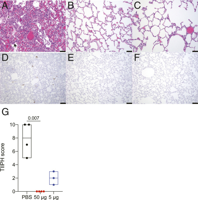
Histopathology and virus detection in the lungs following SARS-CoV-2 challenge. Lung parenchymal tissue was assessed for pathology and viral antigen 7 d postchallenge. (A–C) Paraffin-embedded sections were stained with hematoxylin and eosin and shown for animals that received PBS (A), 50 µg of RFN (B) and 5 μg of RFN (C). Inflammatory debris (white star), type II pneumocyte hyperplasia (black arrow), edema (black triangle), and vasculitis of small- to medium-caliber blood vessels (white arrows) is indicated. (Scale bars, 50 µm.) (D–F) SARS-CoV-2 nucleocapsid detected by IHC in alveolar pneumocytes, pulmonary macrophages, and endothelial cells appears as brown aggregates. (Scale bars, 100 µm.) Representative images are shown. (G) Each pathologic finding was quantified for six lung sections and reported as a combined TIIPH score for all animals necropsied 7 d postchallenge.

