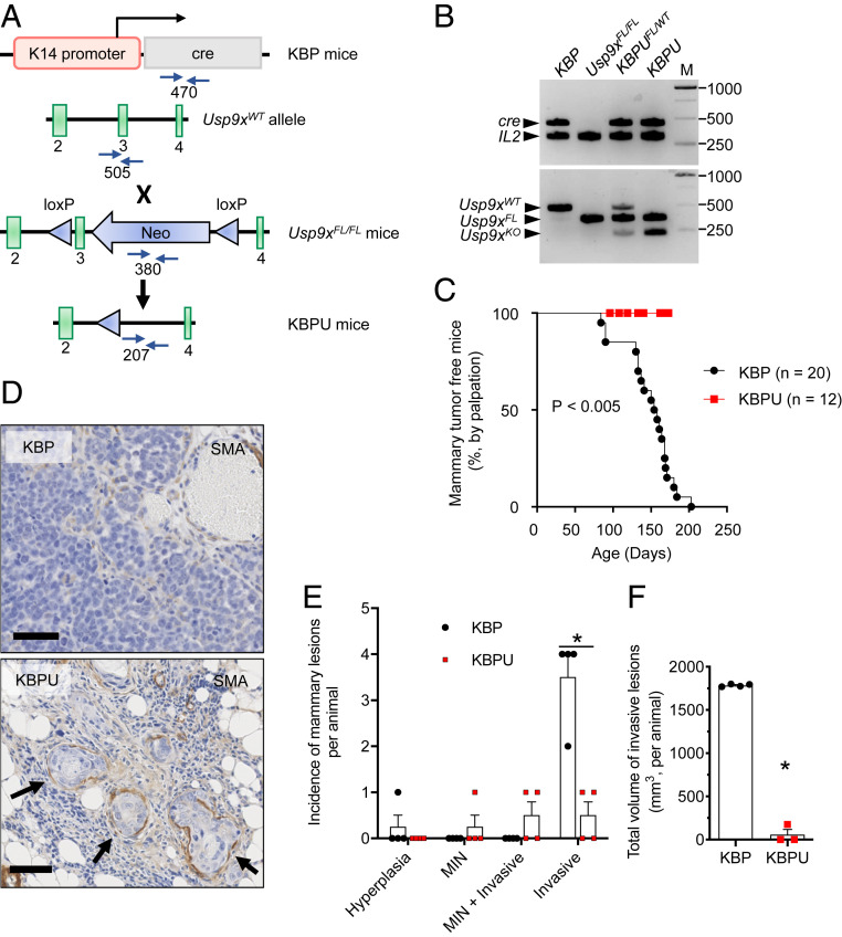
Fig. 5.
USP9x loss of function suppresses tumor progression in a model of spontaneous murine TNBC. (A) Schematic of the generation of KBPU mice via KBP × Usp9xFL/FL cross-breeding. In the conditional allele, loxP sites (blue triangles) flank exon 3 of the endogenous Usp9x locus and the neomycinR cassette (Neo). Primer sites used for genotyping (blue arrows) and the expected PCR product sizes are shown. (B) Confirmation of conditional KO of Usp9x in genomic DNA isolated from tail tissue obtained from KBP × Usp9xFL/FL crosses. Shown is the PCR using primers to detect cre and Il2 (internal control, 324 bp) genes (Top) and the Usp9x wild-type, floxed, and KO alleles (Bottom). KBP and Usp9xFL/FL mice are shown as controls. Note that tail DNA from the KBP × Usp9xFL/FL crosses contains both the Usp9xFL and Usp9xKO alleles resulting from heterogenous K14 expression in tail tissue. M = molecular weight markers in base pairs. (C) Kaplan–Meier analysis showing the latency of palpable mammary tumors in KBP (n = 20 mice) and KBPU mice (n = 12 mice). The P value is determined by the log-rank test. (D) Representative images of anti-SMA–stained sections of mammary glands from KBP and KBPU mice (euthanized at 175 and 173 d of life, respectively). Note that the KBP mammary gland contains a typical invasive mammary carcinoma; the KBPU mammary gland demonstrates mammary intraepithelial neoplasia (MIN) contained within anti-SMA–stained basement membrane (arrows). (Scale bars, 50 µm.) (E) Incidence of mammary lesions per animal at euthanasia (age ∼165 to 180 d, n = 4 mice per group). (F) Total volume of invasive lesions in mammary glands of KBP and KBPU mice from C. Error bars represent SEM. P value is determined by the Student’s t test and corrected for multiple comparisons using the Holm–Sidak method.

