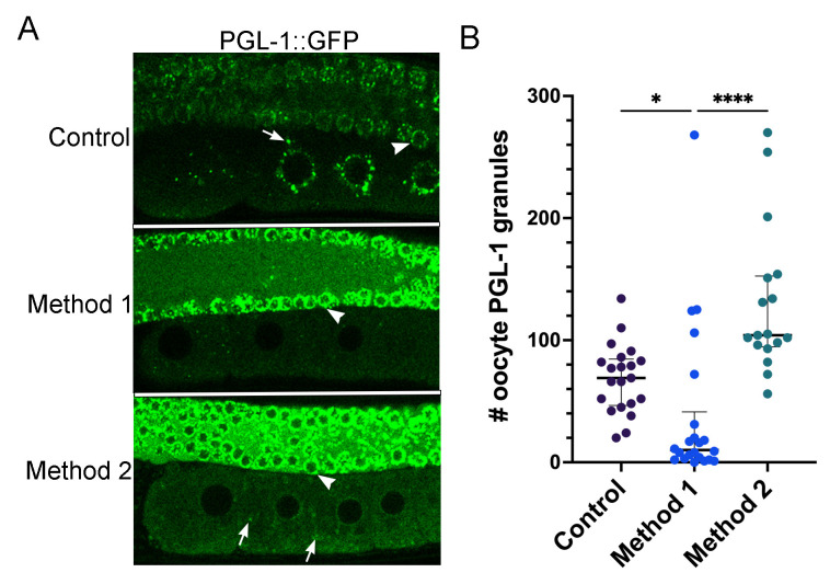
Figure 1. PGL-1::GFP decondenses in oocytes in response to increased heat.
(A) In control, unstressed worms, PGL-1 is condensed into P granules in the proximal oocytes and distal nuclei. Method 1: Worms were incubated at 34°C for 40 min. in a microscope stagetop incubator and imaged at the same temperature. This method resulted in dispersal of PGL-1::GFP in the proximal oocytes of most worms. Method 2: Worms were incubated at 34°C for 40 min. in a standard incubator and imaged at room temperature. This method resulted in significantly more granules than Method 1; the granules were often detached from the nuclear envelope. Arrows indicate granules in oocytes; arrowheads indicate nuclear-associated granules in distal nuclei. (B) Graph shows the number of PGL-1::GFP granules in a single Z-slice of the -2 to -5 oocytes. Error bars represent median with interquartile range, *=P<0.05; ****=P<0.0001.

