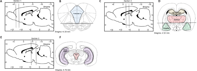
Figure 2.
Schematic depicting rat brain microdissection procedures.
2-mm (sections 1 and 2) or 3-mm thick coronal brain sections (section 3) were cut from either sham-operated or spinal cord injury rats as indicated in (A, C and E) and tissue blocks containing the prefrontal cortex (B), the dorsal hippocampus, thalamus and amygdala (D), or the ventral hippocampus and periaqueductal gray (F) were microdissected under a stereoscopic microscope (magnification 10×) using the Paxinos and Watson rat brain atlas as a reference (Paxinos and Watson, 2006).

