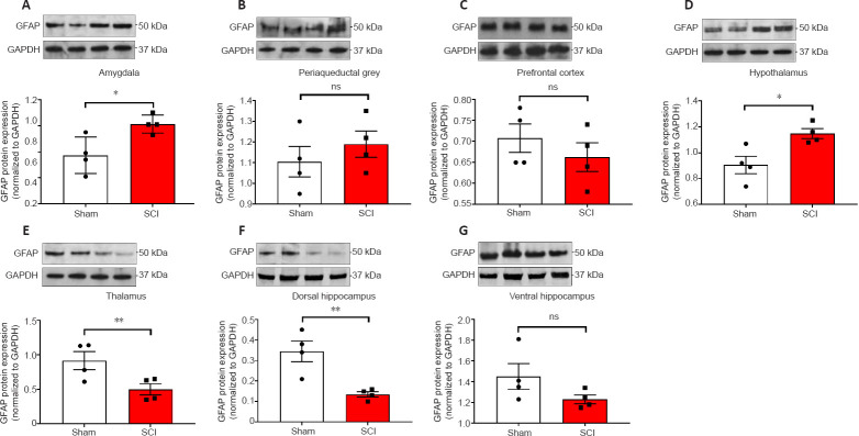
Figure 4.
Western blot analyses of GFAP protein expression in the amygdala, periaqueductal gray, prefrontal cortex, hypothalamus, thalamus, ventral & dorsal hippocampus of sham-injured (Sham) and spinal cord injury rats.
(A–G) Representative GFAP immunoblots and semi-quantitative densitometric analyses are shown for the (A) amygdala, (B) periaqueductal grey, (C) prefrontal cortex, (D) hypothalamus, (E) thalamus, dorsal and ventral hippocampus (F and G). Data are the mean ± SEM of two separate experiments, each run in duplicate. *P< 0.05, **P< 0.01 (Student's t-test). GFAP: Glial fibrillary acidic protein; ns: not significant.

