
Figure 2.
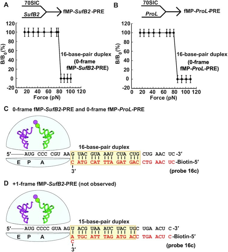
The force profile of fMP-PRE complexes in FIRMS analysis. (A) Formation of an fMP-SufB2-PRE complex by rapid delivery of SufB2-TC to a 70SIC, and (B) formation of an fMP-ProL-PRE complex by rapid delivery of ProL-TC to a 70SIC. A single transition is observed for both (A) and (B), corresponding to dissociation of a 16-base-pair duplex and indicating occupancy of 0-frame of the PRE complex. (C) A scheme representing the immobilized PRE complex in the 0-frame, prior to dissociation of the biotinylated DNA probe, illustrates the sequence of the 16-base-pair mRNA-probe duplex. (D) A scheme representing the immobilized PRE complex in the +1-frame, prior to dissociation of the biotinylated DNA probe, illustrates the sequence of the 15-base-pair mRNA-probe duplex. The duplex up to the entry of the mRNA channel into the ribosome is shaded, while the codons at the A and P site of each complex are marked. Errors were derived from three technical repeats.