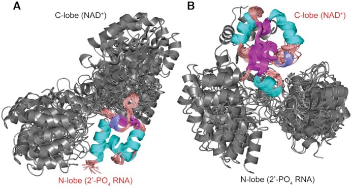
Figure 2.

Structures of the NMR ensemble of apo-RslTpt1 overlaid on the N-lobe (that engages 2′-PO4 RNA, panel A) and the C-lobe (that engages NAD+, panel B). The lobe on which the structures are aligned is labeled in red font. The inability to align the structures on both lobes simultaneously illustrates the fact that while the structures of the individual lobes are well-defined in solution, their relative orientation is not, due to significant inter-lobe flexibility. Given that there are no experimental inter-lobe constraints, the apparent parsing into two sub-families for the unaligned lobe is likely an artifact of the force-field and the limited number of structures (20) selected to represent the NMR ensemble. Elements of secondary structure are indicated with α-helices, β-strands and 310 helices colored cyan, magenta and blue, respectively. Loops are colored salmon.
