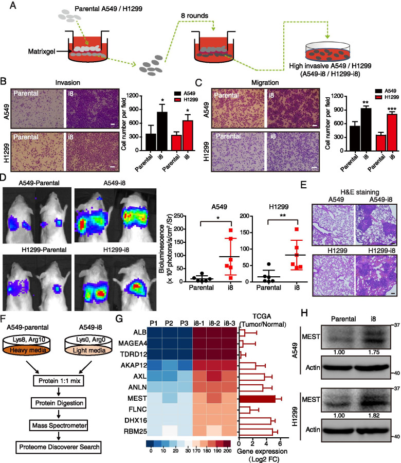
Fig. 1.
SILAC-based proteomics identifies MEST as an invasion regulator in lung cancer cells. A Diagram depicting the establishment of highly invasive cell lines (A549-i8 and H1299-i8) from lung cancer cells via eight rounds of invasion selection. B, C The abilities of invasion (B) and migration (C) of A549-i8 and H1299-i8 cell lines were determined by transwell assays, as compared to their parental cell lines, respectively. Scale bar, 100 μm. D NCG mice were transplanted with luciferase-labeled A549-i8 cells or H1299-i8 cells, or their corresponding luciferase-labeled parental cells (2 × 10⁶ cells per mouse) via tail vein injection (n = 6); mice were then visualized 1 month after transplantation by using an IVIS 200 Imaging System. Lungs harvested after imaging were histologically analyzed by H&E staining (E). Scale bar, 100 μm. F Scheme for the identification of differentially expressed proteins by SILAC-based quantitative proteomics. G The expression levels of the top 10 differentially expressed proteins in A549-i8 cells are represented by heatmap. Their expressions in lung adenocarcinoma and normal tissue were compared by using TCGA dataset (n = 503), showing that MEST is highly expressed in clinical lung cancer tissues, as indicated by red bars. H The expression of MEST in A549-i8 and H1299-i8 cells and in their parental cells was assessed by western blot analysis

