FIGURE 4.

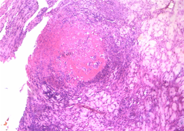
Histological feature of LSD affected nodular lesions. Figure demonstrated deep dermis and subcutis; focal granulomatous lesion comprised of necrotic debris and encircling mononuclear cell infiltration (magnification of image 100×)

Histological feature of LSD affected nodular lesions. Figure demonstrated deep dermis and subcutis; focal granulomatous lesion comprised of necrotic debris and encircling mononuclear cell infiltration (magnification of image 100×)