Figure 1.
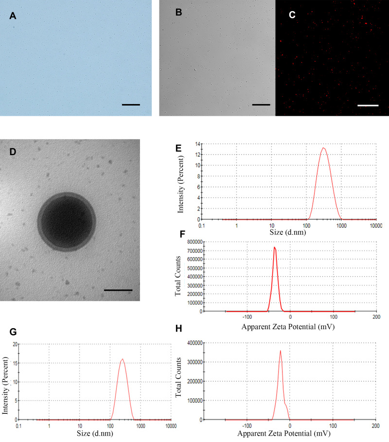
In vitro characteristics of iRGD-ICG-10-HCPT-PFP-NPs and ICG-10-HCPT-PFP-NPs (A) morphology of iRGD-ICG-10-HCPT-PFP-NPs under a light microscope, scale bar: 20 μm (B) morphology of iRGD-ICG-10-HCPT-PFP-NPs under the white light of a CLSM, scale bar: 20 μm (C) morphology of iRGD-ICG-10-HCPT-PFP-NPs under a red fluorescence channel of a CLSM, scale bar: 100 μm (D) structure of iRGD-ICG-10-HCPT-PFP-NPs under TEM, scale bar: 200 nm (E) particle size of iRGD-ICG-10-HCPT-PFP-NPs (F) zeta potential of iRGD-ICG-10-HCPT-PFP-NPs (G) particle size of ICG-10-HCPT-PFP-NPs (H) potential of ICG-10-HCPT-NPs.

