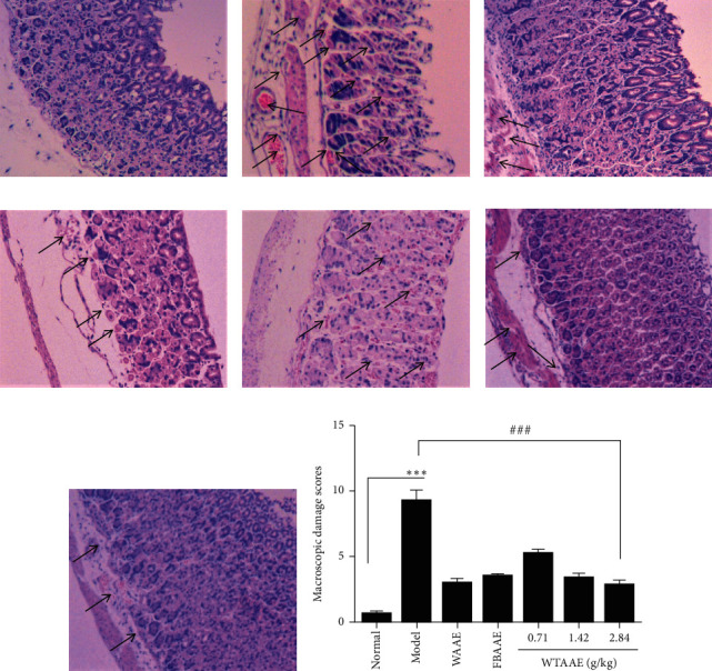
Figure 2.

Effect of the AAEs on gastric histopathology stained by hematoxylin and eosin (H&E). Animals were treated with WAAE (2.84 g/kg), FBAAE (2.84 g/kg), or WTAAE (0.71, 1.42, and 2.84 g/kg) by oral administration. The normal and model groups were given distilled water correspondingly. (a) Normal. (b) Model. (c) WAAE (2.84 g/kg). (d) FBAAE (2.84 g/kg). (e) WTAAE (0.71 g/kg). (f) WTAAE (1.42 g/kg). (g) WTAAE (2.84 g/kg). (h) Pathological damage score analysis from HE staining. Data are expressed as mean ± SD (n = 4). ∗∗∗p < 0.001 vs. normal group; ###p < 0.001 vs. model group.
