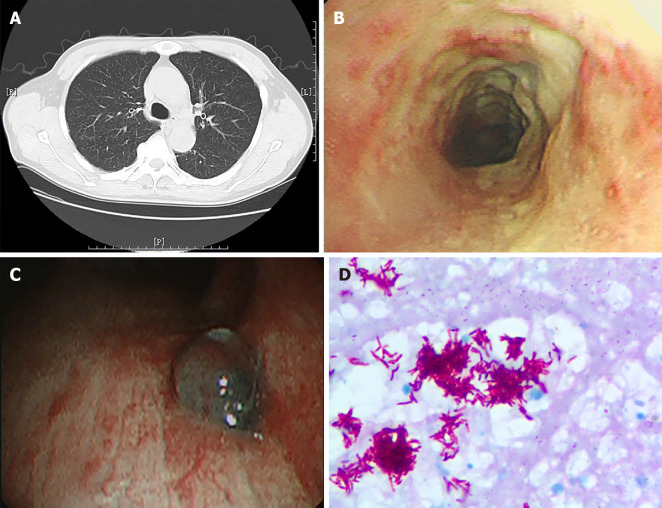
Figure 1.
Chest computed tomography, bronchoscopy image and pathology image of a patient. A: Chest computed tomography of a patient with tracheobronchial tuberculosis showing no obvious abnormalities; B: Bronchoscopy image showing white caseous necrotic tissue on tracheal wall; C: Bronchoscopy image showing granulomatous proliferation on the wall of lower right trachea; D: Necrotic granulomatosis was detected on the pathology image, accompanied with positive acid fast-staining and detection of mycobacterium tuberculosis complex, × 1000 magnification.

