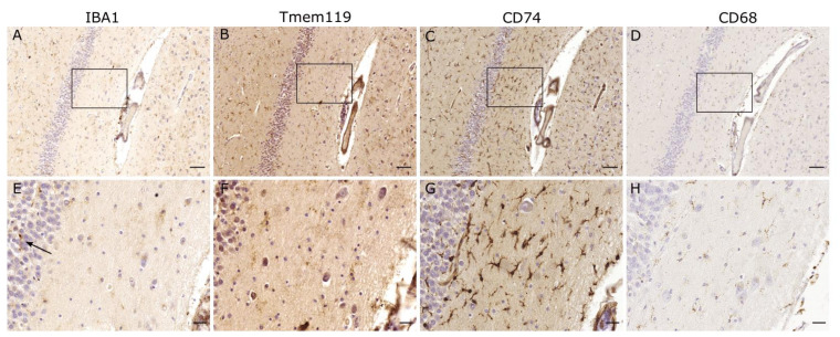
Figure 2.
IBA1-negative microglia. Regions seemingly devoid of microglia in the IBA1 staining, exhibit positive staining for several other microglial markers such as TMEM119, CD74 and CD68. Occurrence of IBA1-positive microglia at the margin (↑) suggests only localized changes. Scale bars: (A–D): 100 µm, (E–H): 25 µm.

