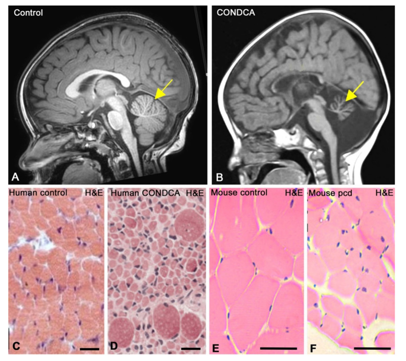
Figure 3.
Cranial magnetic resonance imaging (MRI) of a control (A) and a CONDCA patient (B). Whereas the 20-month-old female healthy control patient (A) shows a typical well-developed cerebellum (yellow arrow), severe cerebellar atrophy (yellow arrow) is observed in the 24-month-old female CONDCA patient (B). (A) Courtesy of Dr. Ana Canga, “Hospital Universitario Marqués de Valdecilla”, Santander (Spain). (B) Adapted from [3]. Copyright 2019 American Journal of Medical Genetics. (C,D) Haematoxylin-eosin (H&E)-stained skeletal muscle tissue biopsies at 7 months of age from healthy control (C) and CONDCA patients (D). Note the fibre atrophy with a few interspersed hypertrophic fibres in the muscle tissue of the patient. Scale bars: 50 µm. Adapted with permission from [1]. Copyright 2018 The EMBO Journal. (E,F) Haematoxylin-eosin (E,H)-stained skeletal muscle tissue cross-sections from control (E) and pcd (F) mice. Note the muscle atrophy and the notable reduction in muscle fibre size in the pcd mouse. Scale bars: 30 µm. Adapted with permission from [37]. Copyright 2018 Journal of Tissue Engineering and Regenerative Medicine.

