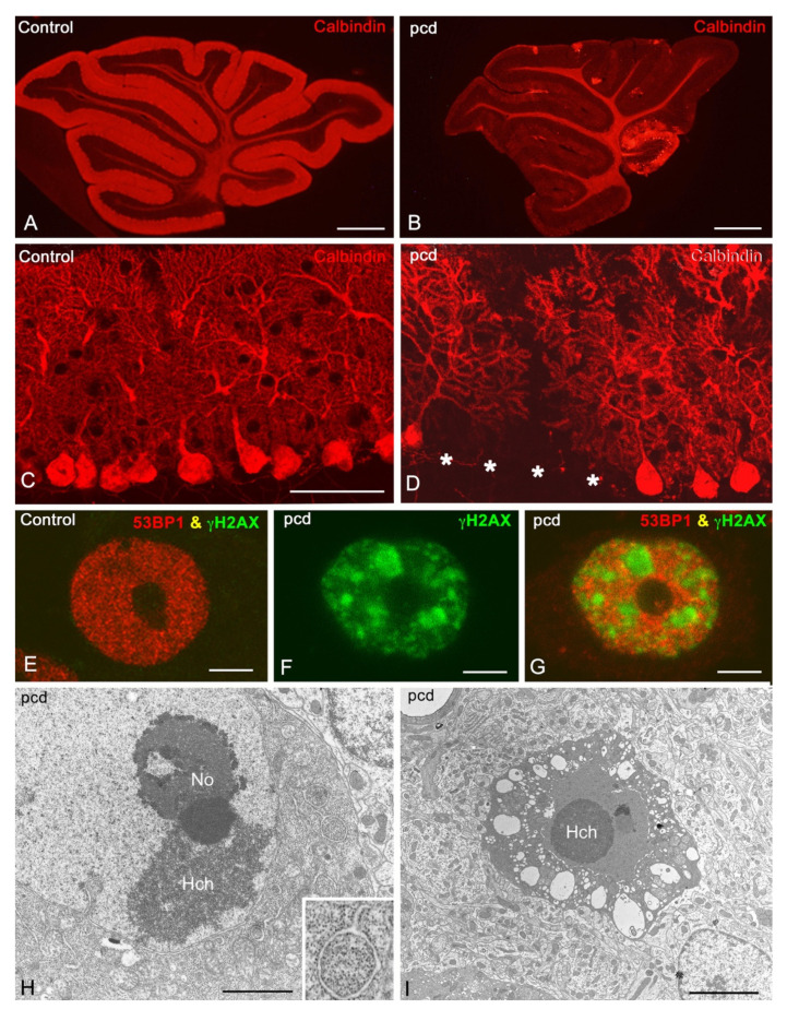
Figure 4.
Degenerative features of the Purkinje cells in the pcd mice. (A,B) Representative confocal microscopy images of sagittal sections of the vermis of P30 control (A) and P30 (B) pcd mutant mice immunolabelled for calbindin D-28k. Note that in the pcd mouse there was a dramatic reduction in calbindin immunostaining in the molecular and PC layers resulting from the massive loss of PCs at P30 and that only PCs located in lobule X remained (arrow in B). Scale bars: 1 mm. (C,D) High magnification of calbindin D-28K immunolabelling of PC perikarya and their dendritic trees in control (C) and pcd mice (D) at P20. Note the loss of PCs (white asterisks) in the pcd mouse. Scale bar: 100 µm. (E–G) Confocal microscopy images of PC nuclei from control (E) and pcd mice (F,G) at P20 double immunolabelled for the modified histone γH2AX (green), a marker of DNA double-strand breaks at sites of DNA damage, and p53-binding protein 1 (P53BP1, red), a key DNA repair factor. (E) Note the absence of γH2AX labelling and the typical diffuse nucleoplasmic distribution of 53BP1, in the control PC nucleus, excluding the nucleolus. (F,G) In contrast, the nucleus of the pcd mouse shows prominent nuclear foci of DNA damage immunostained for γH2AX (F). Although the DNA repair factor 53BP1 was expressed in the nucleoplasm, it was not concentrated in γH2AX-positive nuclear foci of DNA lesions (G), indicating defective DNA repair. Scale bars: 5 μm. (H) Electron microscopy image of PCs from pcd mice at P20. Free polyribosomes were replaced by densely packed monoribosomes. Cytoplasmic portions containing monoribosomes appear sequestered in autophagic vacuoles bound by isolated RE cisternae (insert). Scale bar: 1 μm. (I) Electron microscopy image of mutant apoptotic PC Scale bars: 5 μm. (A,B) Adapted with permission from [17]. Copyright 2011 The Journal of Biological Chemistry. (C,D,H) Adapted with permission from [48]. Copyright 2011 Brain Pathology. (E–G) Adapted with permission from [49]. Copyright 2019 Neurobiology of Disease.

