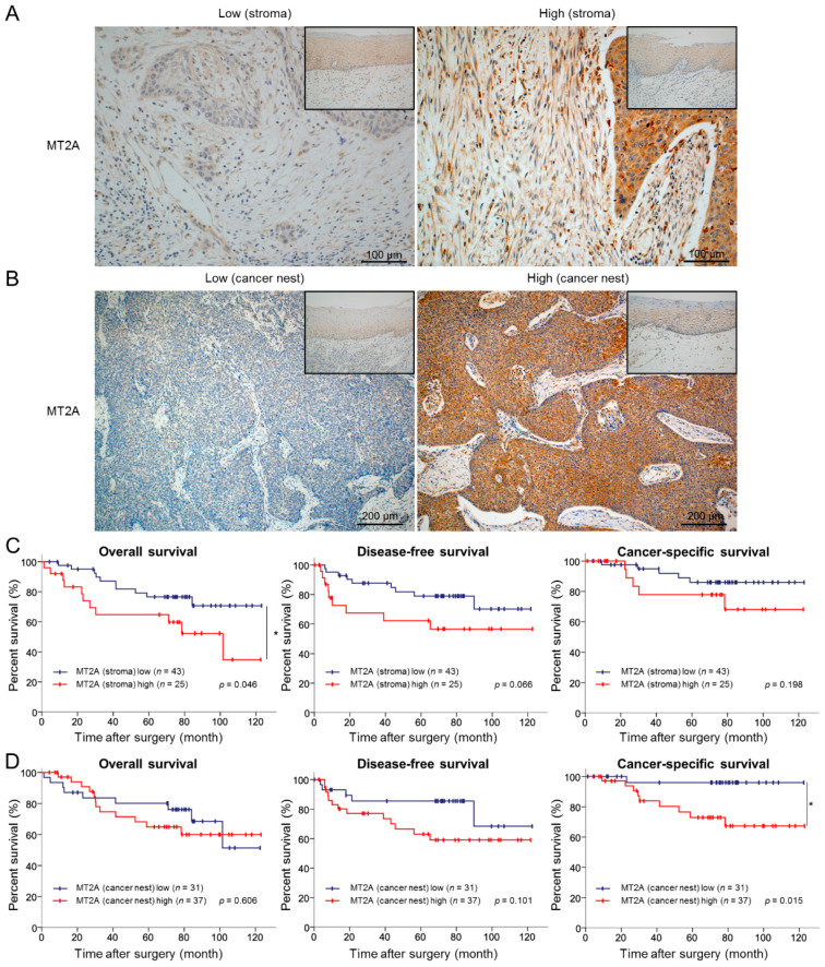
Figure 6.
High expression levels of MT2A in the cancer stroma and cancer nest correlated to a poor prognosis of ESCC patients. (A) Immunohistochemical staining for MT2A in the cancer stroma of 69 human ESCC tissues. Representative immunostaining images show low intensity (left) and high intensity (right) with corresponding normal esophageal squamous epithelia (insets). Scale bars = 100 μm. (B) Immunohistochemical staining for MT2A in the cancer nest of 69 human ESCC tissues. Representative immunostaining images show low intensity (left) and high intensity (right) with corresponding normal esophageal squamous epithelia (insets). Scale bars = 200 μm. (C) Kaplan–Meier curves for overall survival, disease-free survival, and cancer-specific survival in 68 ESCC patients stratified into two groups based on MT2A expression levels in the cancer stroma: MT2A low cases (n = 43) and MT2A high cases (n = 25). (D) Kaplan–Meier curves for overall survival, disease-free survival, and cancer-specific survival in 68 ESCC patients stratified into two groups based on MT2A expression levels in the cancer nest: MT2A low cases (n = 31) and MT2A high cases (n = 37). Data are analyzed by log-rank test (* p < 0.05).

