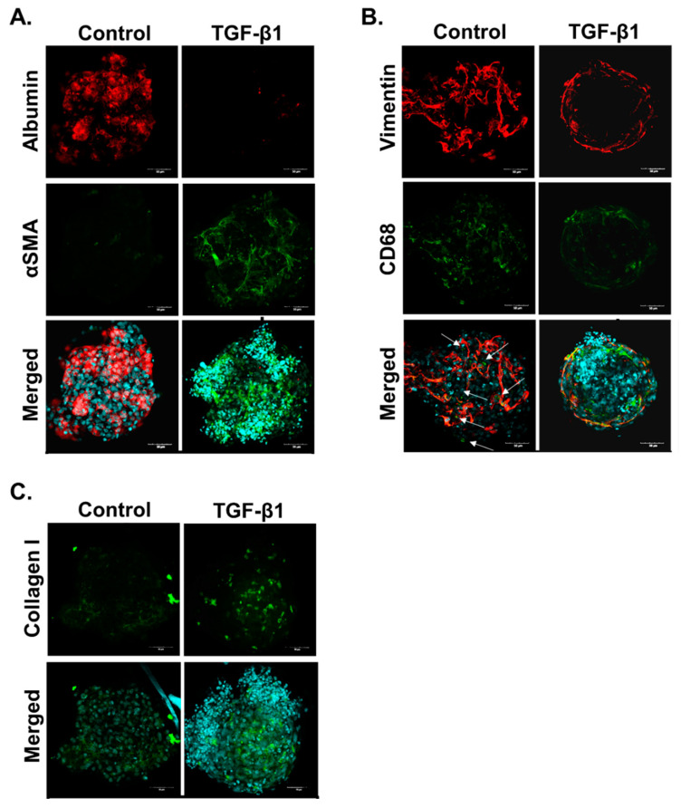
Figure 2.
TGF-β1 induces fibrosis in human liver MTs. MTs were left untreated or exposed to 1 ng/mL TGF-β1 for 10 days which was refreshed every 2–3 days. The MTs were fixed and stained to demonstrate localization of the three cell types: HepaRG cells stain positive for albumin; hTERT-HSC stain positive for αSMA and vimentin; and THP-1 stain positive for CD68 and vimentin. HSC activation and ECM deposition were shown through increased αSMA and collagen I staining, respectively. Images are shown as a maximum intensity projection including the merged image with DAPI for each staining combination: albumin and αSMA (A), vimentin and CD68 (B) and collagen (C). Arrows identify THP-1 as these cells are positive for both CD68 and vimentin (B).

