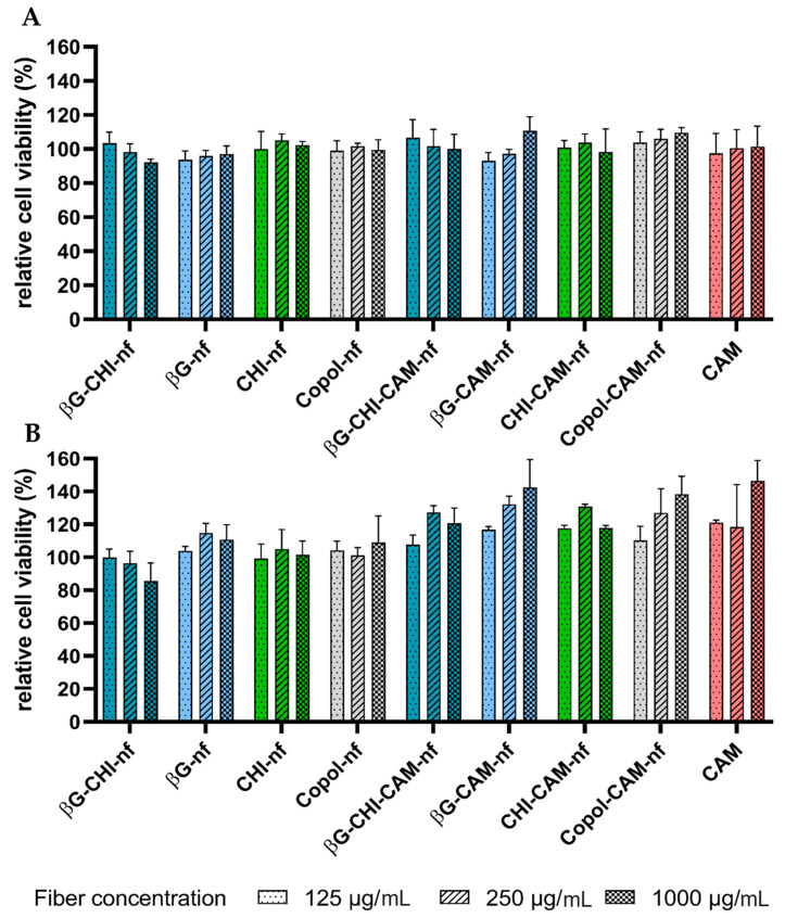
Figure 3.
Relative cell viability (%) of (A) HaCaT cells and (B) macrophages (RAW 264.7) after 24 h incubation at 37 °C and exposure to nanofibers (dissolved in concentrations of 125, 250 and 1000 µg/mL) and chloramphenicol (CAM) (in concentrations of 1.25, 2.5 and 10 µg/mL). Results are presented as mean ± SD (n = 3). Abbreviations: βG (β-glucan), CHI (chitosan), Copol (co-polymers: polyethylene oxide and hydroxypropylmethylcellulose), CAM (chloramphenicol), nf (nanofiber).

