Figure 1.
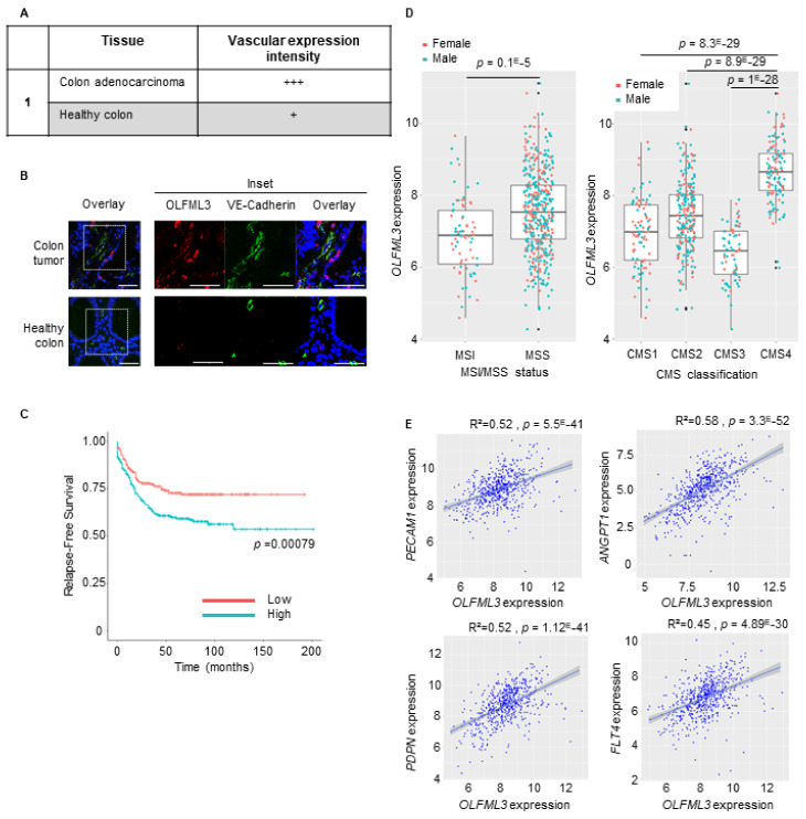
Expression of OLFML3 in colorectal cancer (CRC) correlates with shorter relapse-free survival, with microsatellite stable status (MMS) and consensus molecular subtypes 4 (CMS4), and with the expression of angiogenic factors. (A) Vascular expression intensity of OLFML3 was ranked as follows: none (−), low (+), moderate (++), or strong (+++). (B) Representative confocal images of VE-cadherin+ (green)- and OLFML3+ (red)-stained tumor vasculature in sections of CRC and healthy colon tissue. Note the difference in abundance of vascular VE-cadherin+ and perivascular cells in OLFML3-expressing tumors relative to that in healthy colon tissue. DAPI (blue), nuclear counterstain (overlays). Scale bars = 50 μm. The right panel is a zoom image of the frame of the left panel. (C) Kaplan–Meier survival analysis of the index of OLFML3 mRNA expression (low (red) vs. high (blue)) relative to the median value in CRC patient tumor samples. ((D), left) Expression of OLFML3 mRNA in CRC patient tumor samples according to microsatellite instable/microsatellite stable status (MSI/MSS). ((D), right) Expression of OLFML3 mRNA in four consensus molecular subtypes (CMS 1–4) from CRC patients. (E) Scatter plots with linear regression lines (blue) showing the correlation between levels of OLFML3 mRNA and that of angiogenic markers (PECAM1, ANGPT1, PDPN, FLT4) in CRC samples and normal colon tissue. Levels of OLFML3 mRNA in different subgroups of patients were compared using a t-test with equal variance. The correlation between the expression of OLFML3 and that of genes of interest was calculated using Pearson’s and Spearman’s rank correlation analyses.

